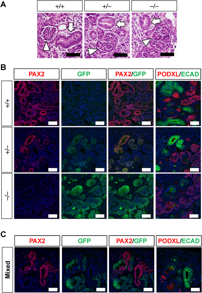
Figure 2.

Successful deletion of PAX2 in renal tubules derived from iPS cells. (A) Hematoxylin–eosin (HE) staining of kidney tissues induced from wild-type (+/+), heterozygous (+/−), and homozygous (−/−) clones. Arrowheads: glomeruli; arrows: renal tubules. (B) PAX2 (red) and GFP (green) staining of kidney tissues induced from the three genotypes. Rightmost columns: neighbouring sections were stained with markers for renal tubules (E-cadherin: ECAD) and glomerular podocytes (podocalyxin: PODXL). (C) Nephron formation from a mixed colony of iPS cells. Nephron epithelia are composed of PAX2+ wild-type and GFP+ mutant cells. Scale bars: 50 µm.
