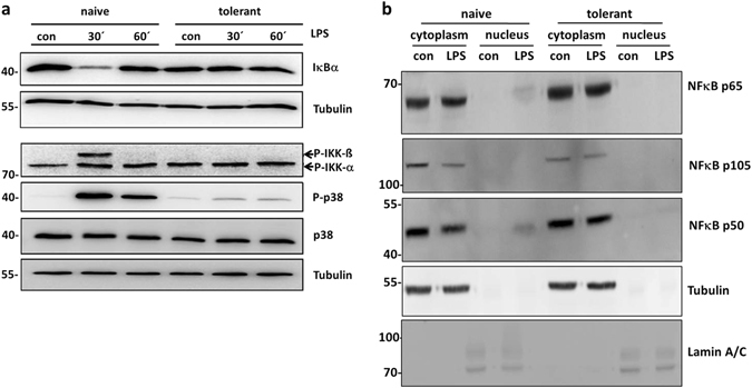
Figure 4.

Impaired TLR4 signalling in tolerant BMMCs. Cells were preincubated with LPS (300 ng/ml) for 24 h to induce ET and stimulated with LPS (1 µg/ml) for 30 min and 60 min. Subsequently, cells were lysed in the presence of phosphatase inhibitors. SDS-PAGE and Western blotting with antibodies specific for IκBα, P-IKK-α/-β, P-p38, p38 and Tubulin (loading control) was performed (a). Because proteins of comparable size (p38 and IκBα) were analysed, the same lysates were separated on two gels with an anti-Tubulin loading control on each gel. Moreover, naive and tolerant cells were stimulated with LPS (2 µg/ml) for 60 min and a cytosol/nucleus fractionation was performed, followed by subsequent SDS-PAGE and Western blotting with antibodies specific for NFκB p65, NFκB p105 and p50 (both are detected by the same antibody), Tubulin, and Lamin A/C as loading controls (b). Western Blots are representative of two independent experiments with two biological samples.
