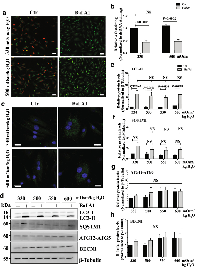
Figure 5.

Hyperosmolarity does not influence autophagic flux in NP cells. (a) Acridine orange staining of NP cells cultured under iso- (330 mOsm/kg H2O, top row) or hyperosmotic (500 mOsm/kg H2O, bottom row) condition, treated with (right) or without (left) bafilomycin A1. Hyperosmotic stimulus alone showed no change in number of acidic organelles. Bafilomycin A1 significantly reduced the acridine orange staining irrespective of osmolarity. Scale bar, 35 μm. (b) Quantification of acridine orange staining confirmed that hyperosmolarity had no effect on the number of acidic organelles. (c) LC3 immunofluorescence staining of NP cells cultured under hyperosmolarity with or without bafilomycin A1 treatment. Hyperosmotic stimulus did not upregulate LC3-positive autophagosomes. Scale bar, 20 μm. (d) Western blot analysis of NP cells cultured under increasing osmolarity (330–600 mOsm/kg H2O) with or without bafilomycin A1 treatment. The accumulation of LC3-II with bafilomycin A1 treatment was similar under iso- and hyperosmotic conditions. SQSTM1 also showed a trend of accumulation with bafilomycin A1 treatment under all conditions. The levels of ATG12-ATG5 and BECN1 remained unaltered between the experimental groups. (e–h) Densitometric analyses of multiple Western blots shown in (d). Bars represent mean ± SEM (n = 5). Two-way ANOVA with Tukey’s multiple comparisons test was used to determine statistical significance. NS, non-significant. BafA1, bafilomycin A1. Western blot images were cropped and acquired under same experimental conditions. See Supplementary Fig. S1 for examples of uncropped images.
