
Figure 8.
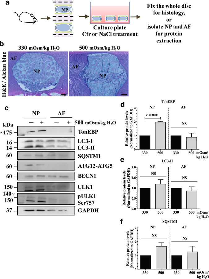
NP cells do not induce autophagy in response to hyperosmotic stimulus in an ex vivo disc organ culture model. (a) A schematic depicting ex vivo rat intervertebral disc organ culture model. (b) H&E and alcian blue staining of discs cultured under iso- (330 mOsm/kg H2O) or hyperosmotic (500 mOsm/kg H2O) conditions showing that NP maintained its structure and cellular morphology. Scale bar: 100 μm. (c) Western blot analysis of tissue proteins from NP or AF (annulus fibrosus) compartments of the organ culture discs. The level of TonEBP increased with hyperosmotic stimulus only in the NP. However, the levels of LC3-II, SQSTM1, ATG12-ATG5, BECN1, as well as pULK1 Ser757 did not change with hyperosmolarity in both NP and AF. (d–f) Densitometric analyses of multiple Western blots represented in (c). Bars represent mean ± SEM (n = 3; For each independent experiment, one motion segment per group was used for histology and 6 motion segments per group were used for tissue protein Western blot). Student t test was used to determine statistical significance. NS, non-significant. Western blot images were cropped and acquired under same experimental conditions. See Supplementary Fig. S1 for examples of uncropped images.