
Figure 7.
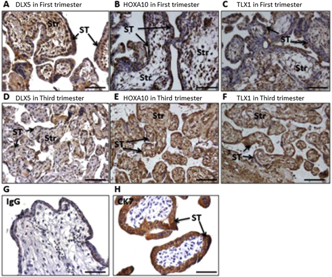
Localisation of DLX5, HOXA10 and TLX1 proteins in first and third trimester placentae Representative sections of n = 5 first trimester and n = 5 third trimester placentae. DLX5, HOXA10 and TLX1 are expressed in the syncytiotrophoblast layer (black arrows) of first trimester (A–C) and third trimester (D–F) placentae. CK7 was used as a positive marker (H) and IgG as a negative control (G). (I) Dot plot shows immunoreactive signal pixel density for DLX5, HOXA10 and TLX1 in first and third trimester villi. Intensity for all three candidate proteins was significantly lower in the third trimester. The intensity of staining was calculated using an AxioVision software (Carl Zeiss KS 400, Germany) from five fields of view of each placental section and the results were expressed as the mean pixel density (in arbitrary units). Str = stromal.