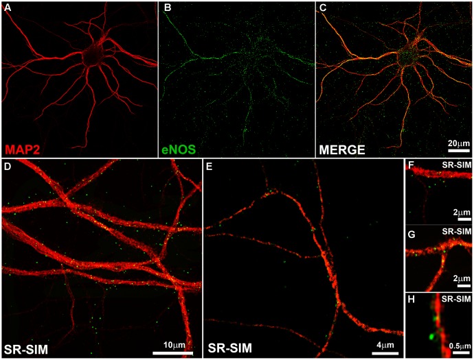
FIGURE 1.
The endothelial isoform (eNOS) puncta decorate the dendritic tree. (A–C) Confocal microscopy shows the dendritic marker MAP2 (red) and a punctate eNOS pattern (green) in cortical cells. (D–E) SR-SIM microscopy in hippocampal cells using the same antibodies. (F–H) SR-SIM images of dendrite segments of hippocampal cells at higher magnification. The calibration bars in the corresponding panels are indicated.

