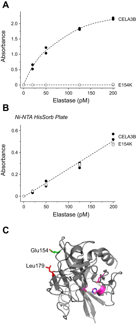
Fig. 6.

Detection of CELA3B mutant E154K by the ScheBo Pancreatic Elastase 1 Stool Test. A: purified wild-type and E154K mutant CELA3B were assayed at 20–200 pM. B: purified wild-type and E154K mutant CELA3B were immobilized through their COOH-terminal His tags to Ni-NTA HisSorb plates (Qiagen) and detected with the biotinylated antibody-streptavidin-peroxidase complex from the ScheBo test. Assays were performed in duplicate, and both data points were plotted. C: structural model of human CELA3B indicating the positions of Glu154 (green) and nearby Leu179 (red). Also shown for reference are residues of the catalytic triad (magenta). Structural model for active CELA3B was generated by the SWISS-MODEL protein structure homology-modeling server (2) with porcine elastase used as template (PDB file 3UOU). Image was rendered with PyMOL (Schrödinger, Cambridge, MA).
