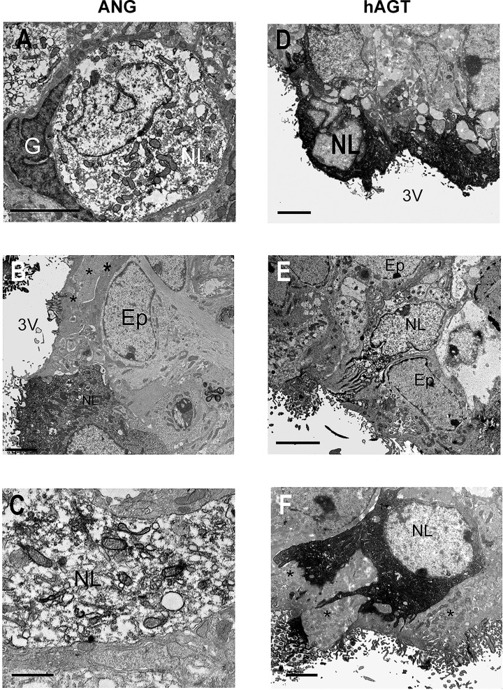
Fig. 2.
Electron microscopic localization of ANG peptides and hAGT in SFO. Ultrastructural appearance of ANG peptide (A–C) and hAGT (D–F) cell immunoreactivity in the SFO in NT (A and B), A+ (E), and SRA (C, D, and F) mice. ANG peptide immunoreactivity was fairly sparsely distributed in the perikarya (A) and dendrites (C) of neuron-like (NL) cells in both NT (A) and transgenic (C) animals. In contrast, more superficially located neuron-like cells exhibited stronger immunoreactivity (D and F). Immunoreactive cell processes could be observed passing between ependymal cell (Ep) processes (* in F) to the surface of the third ventricle (3V). Note the absence of ANG peptide and hAGT immunoreactivity in glia (G) and Ep cells adjacent to immunoreactive NL cells (A, B, and F). Scale bars A and C–F = 2 μm; scale bar B = 0.5 μm.

