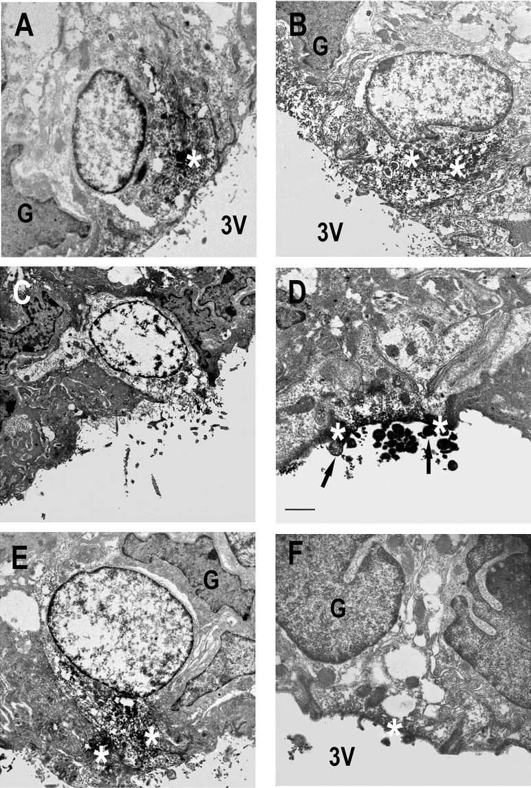
Fig. 5.
Localization of ANG and AGT immunoreactivity near the ventricular surface. Superficial cells of SFO from NT (A and B), A+ (C), and SRA (D–F) mice showing accumulation of ANG peptide (A and B) and hAGT (C–F) immunoreactivity (white *) at the ventricular surface after 35 min of CSF withdrawal. The presence of reaction product in microvilli (arrows in D) is clearly shown. Scale bars in A–C and E = 2 μm; scale bar in D = 0.5 μm; scale bar in F = 1 μm.

