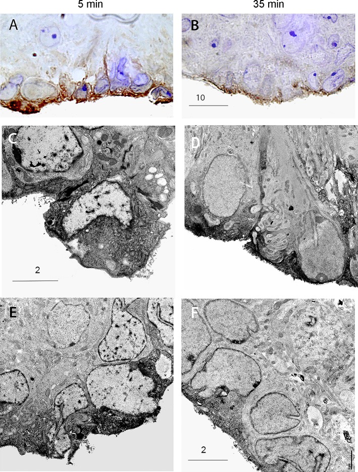
Fig. 6.
Immunocytochemical detection of synaptobrevin-2 (VAMP2) in SFO. Immunocytochemical detection of VAMP2 immunoreactivity in SFO after 5 min (A, C, and E) and after 35 min (B, D, and F) of CSF withdrawal. Note the shift in VAMP2 immunoreactivity to the surface following 35 min of CSF withdrawal and the similar morphology of immunoreactive cells to those in Figs. 2 and 5. Scale bars are as indicated in the figure (in μm).

