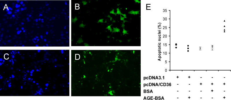
Figure 6. Expression of CD36 Transgene Confers Susceptibility to AGE-BSA-Induced Apoptosis.
(A–D) Representative images show DAPI (A and C) and FITC (B and D) labeling of CD36-negative MCT cells treated with 40 μM AGE-BSA5 for 24 h after co-transfection with green fluorescent protein plasmid pEGFP and pcDNA3.1 empty control vector (A and B), or pEGFP and CD36 expression plasmid pcDNA3.1/CD36 (C and D).
(E) The dot plot shows results of four independent experiments where apoptotic nuclei per 100 total cells were quantitated in transfected cell cultures with or without treatments as indicated.

