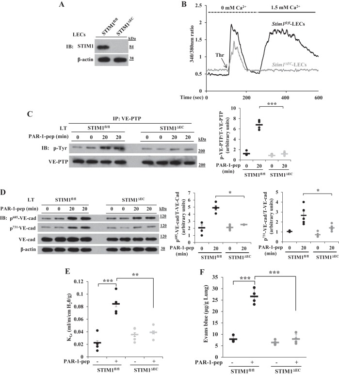
Fig. 2.
Protease-activated receptor 1 (PAR-1)-induced Ca2+ entry, phosphorylation of VE-PTP, phosphorylation of VE-cadherin, and increase in lung vascular permeability are impaired in Stim1ΔEC mice. A: lung endothelial cells (LECs) from Stim1fl/fl and Stim1ΔEC mice were used for immunoblot to determine STIM1 expression. B: LECs from Stim1fl/fl and Stim1ΔEC mice were used to measure thrombin-induced ER-store Ca2+ release and Ca2+ release-activated Ca2+ entry. Data in A and B are representative of 3 independent experiments. C: Stim1fl/fl and Stim1ΔEC mice were intravenously injected with PAR-1 agonist peptide (see materials and methods). 0 and 20 min after administration of PAR-1 agonist peptide, lungs were harvested, and lung tissue extracts were prepared for IP with VE-PTP pAb and blotted with phosphotyrosine mAb. D: lung tissue extracts from Stim1fl/fl and Stim1ΔEC mice were immunoblotted with the indicated phospho-tyrosine-specific VE-cadherin antibodies. Quantified results are presented as the ratio of phosphorylated:total protein in C and D. n = 4 mice for each group. *P < 0.05; ***P < 0.001; Stim1fl/fl vs. Stim1ΔEC. E: lungs from Stim1fl/fl and Stim1ΔEC mice were isolated to determine lung vascular permeability (see materials and methods). PAR-1 agonist peptide (25 μM) was included in the perfusion buffer to determine PAR-1-induced microvessel filtration coefficient (Kf,c). n = 4 mice per group; **P < 0.01; ***P < 0.001; control Stim1fl/fl vs. PAR-1 peptide Stim1fl/fl or Stim1fl/fl PAR-1 peptide vs. Stim1ΔEC PAR-1 peptide. F: Stim1fl/fl and Stim1ΔEC mice received PAR-1 agonist peptide intravenously and were subsequently used to measure Evans blue dye conjugated with albumin (EBA) uptake in the lungs. Results are means ± SE n = 4 mice per group; ***P < 0.001; control Stim1fl/fl vs. PAR-1 peptide Stim1fl/fl or Stim1fl/fl PAR-1 peptide vs. Stim1ΔEC PAR-1 peptide.

