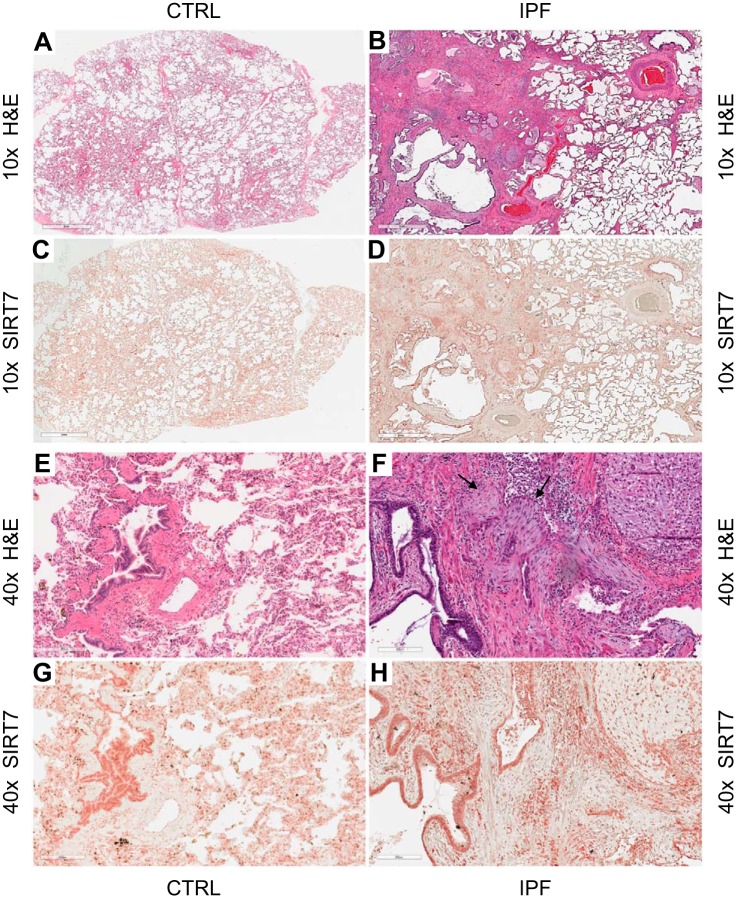
Fig. 4.
Histologic and immunohistochemical analyses of lung tissues. Serial lung sections from a healthy donor (CTRL; A, C, E, and G) and from a patient with IPF (B, D, F, and H) were stained with hematoxylin and eosin (H&E) or SIRT7, as indicated. Digital images were acquired with ×10 (A–D) or ×40 (E–H) objectives. SIRT7-positive cells stain brown, with greater intensity in the nucleus. These experiments were repeated in three healthy control subjects and six patients with IPF with similar results. Arrows in F indicate fribroblastic foci.

