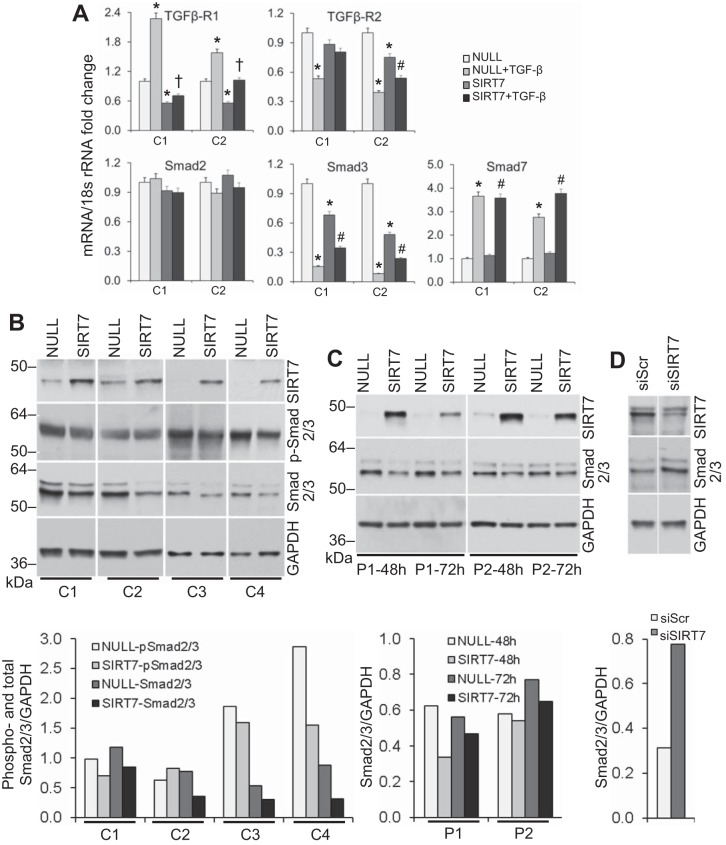
Fig. 8.
Effect of SIRT7 overexpression on TGF-β pathway-related molecules. A: RT-qPCR for Smad2, Smad3, Smad7, TGFβ-R1, and TGFβ-R2 in cultured primary pulmonary fibroblasts from two healthy controls (C1 and C2) transfected with NULL or SIRT7 plasmids and stimulated with rhTGF-β1 48 h later. Analyses were performed 24 h after TGF-β stimulation. *Significant difference (P < 0.05) from NULL-transfected cells without TGF-β stimulation. †Significant difference from NULL-transfected cells with TGF-β. #Significant differences from SIRT7-transfected cells without TGF-β. Note the decreases in SMAD3 and TGFβ-R1 in fibroblast cultures overexpressing SIRT7. B: changes in the expression levels of phosphorylated and total Smad2/3 measured by Western blot analysis in cultured primary lung fibroblasts from four different healthy controls (C1–C4) following overexpression of SIRT7. Fibroblasts were electroporated with a control noncoding (NULL) or SIRT7-encoding plasmid, and Western blots for the indicated targets were performed 72 h later. Noncontiguous gels are demarcated by white spaces. B, bottom: densities for phosphorylated and total Smad2/3 normalized to GAPDH are shown. C: changes in total Smad2/3 levels at indicated times in two patients with IPF (P1, P2) following overexpression of SIRT7. The SIRT7 and GAPDH bands for patient 1 are the same as those for the patient shown in Fig. 7D. C, bottom: total Smad2/3 densities normalized to GAPDH are shown. Note the decreases in Smad2/3 levels in both normal and patient SIRT7-overexpressing cultures. D: changes in total Smad2/3 levels in NHLF following SIRT7 silencing with siRNA. Fibroblasts were electroporated with scrambled (Scr) or SIRT7 siRNA (siSIRT7), and analyses were performed 72 h later. Noncontiguous gels are demarcated by white spaces. SIRT7 and GAPDH bands are the same as those for donor 2 at 72 h shown in Fig. 6A. D, bottom: Densities for total Smad2/3 relative to GAPDH are shown.

