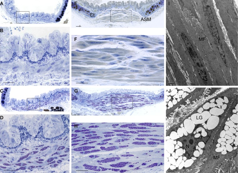
Fig. 1.
Glycogen accumulation in airway smooth muscle of Gaa−/− mice. A–H: representative plastic-embedded 2-µm cross sections stained using the periodic acid Schiff (PAS) method (purple) and counterstained with toluidine blue. Shown are illustratrations of wild-type (WT) bronchi (A and B) and trachea (E and F) (B and F are enlargements of A and E, respectively). Shown are Gaa−/− bronchi (C and D) and trachea (G and H) (D and H are enlargements of C and G, respectively). I and J: low-magnification electron micrographs of airway smooth muscle of WT (I) and Gaa−/− (J) mouse trachealis muscle cells. C, cartilage; ASM, airway smooth muscle cells; MF, myofilaments; LG, lysosomal glycogen; N, nuclei. Scale bar = 50 µm (A, C, E, and G), 25 µm (B, D, F, and H), and 2 µm (I and J).

