
Figure 4.
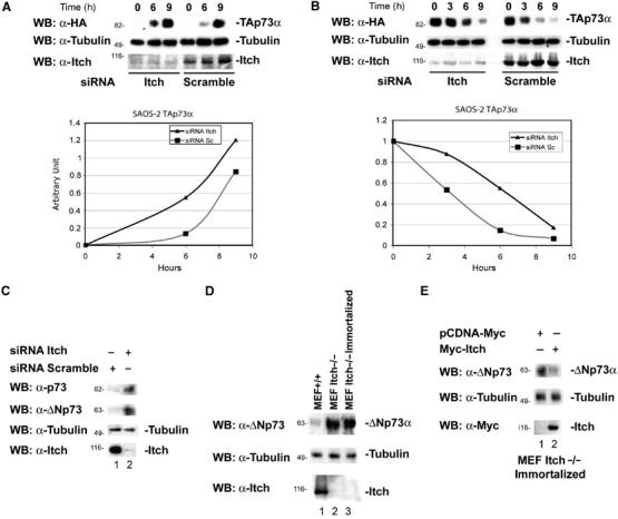
Effects of Itch downregulation on p73 protein levels. (A) Saos-2-TAp73α inducible cells were transfected with siRNA oligonucleotides targetting the Itch sequence or with a scrambled oligonucleotide. After 48 h, cells were induced to express TAp73α for the indicated time points with doxycycline (inducer). p73 levels increase more rapidly and reach higher levels when Itch is downregulated. The lower panel shows endogenous Itch levels. Graphs show densitometric analysis of the p73 western blots normalized for β-Tubulin. (B) Saos-2-TAp73α-inducible cells were transfected with oligos targetting the Itch sequence, or with a scrambled oligo. Cells were induced to express TAp73α for 14 h with doxycycline, the inducer was removed and cells collected at the indicated time points. p73 levels decay more rapidly in cells transfected with the scrambled oligo compared to those in which Itch is downregulated. The lower panel shows endogenous Itch levels. Graphs show densitometric analysis of the p73 western blots normalized for β-Tubulin. (C) Saos-2 cells were transfected with oligos targeting the Itch sequence or with a scrambled oligo and collected 48 h later. Itch downregulation (lower panel) results in an increase of TA and ΔN p73a levels (upper panels). (D) Western blot for endogenous p73 of WT MEFs (MEF+/+), non-agouti-lethal 18H Itch-deficient MEFs (MEF Itch−/−) and a spontaneously immortalized clone of these MEFs (MEF Itch−/− Immortalized). ΔNp73 levels (the only form detectable in these cells) are higher in MEFs Itch−/−. (E) Immortalized MEFs Itch−/− were transfected with Myc-Itch WT and collected 48 h later. Re-introduction of Itch results in ΔNp73 downregulation.