
Figure 3.
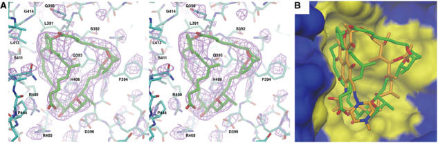
Sor-RNAP cocrystal structure and comparison with Rif. (A) Stereo view of Sor in its binding pocket of Taq core RNAP. Atoms are color-coded as follows: carbon atoms of the RNAP β subunit, cyan; carbon atoms of Sor, green; oxygen, red; nitrogen, blue; sulfur, yellow. Electron density, calculated using (∣FoSor−Fonat∣) coefficients (Sor denotes the Sor-RNAP cocrystal, nat denotes the native core RNAP crystal), is shown in magenta (contoured at 3σ), and was computed using phases from the native RNAP model. Selected amino-acid residues discussed in the text are labeled. (B) View of the antibiotic binding pocket of the RNAP β subunit (same view as A). The RNAP is shown as a surface view, with the β subunit colored blue, but with residues within 4 Å of Sor colored yellow (to define the antibiotic binding pocket). Superimposed in the binding pocket are the structures of Sor (green carbon atoms) and Rif (orange carbon atoms).