
Figure 4.
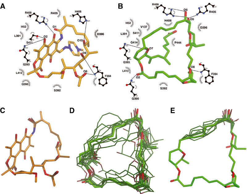
Rif and Sor interactions with RNAP, and conformational flexibility. (A, B) Schematic drawing of RNAP β subunit interactions with Rif (A) and Sor (B), modified from LIGPLOT (Wallace et al, 1995). Residues forming van der Waal's interactions are indicated; those participating in hydrogen bonds are shown in a ball-and-stick representation, with hydrogen bonds depicted as lines. (C–E) Results of molecular dynamics simulations. Starting conformations of Rif (C) or Sor (D, E) are shown as thick bonds and colored light orange (Rif) or light green (Sor). The final conformations after 10 independent molecular dynamics simulations are shown as thinner bonds and darker color. In (E), only atoms C22–C30/O5–O6 were allowed to move during the simulation. In all of the images, flexible, branched segments of each antibiotic that do not interact with RNAP (C38–C43/N2–N4 for Rif, C36–C45/O10–O11 for Sor) were included in the simulations, but were not included in the alignment and are not shown.