
Figure 3.
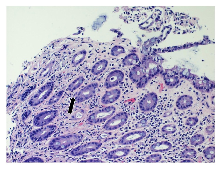
Colonic ulcer biopsy demonstrating ischemic morphology with hyalinization of the lamina propria and atrophic crypts. Small cystic forms are evident. The arrows refer to toxoplasmosis cyst present within the colonic ulcer biopsy.
Official websites use .gov
A
.gov website belongs to an official
government organization in the United States.
Secure .gov websites use HTTPS
A lock (
) or https:// means you've safely
connected to the .gov website. Share sensitive
information only on official, secure websites.
Colonic ulcer biopsy demonstrating ischemic morphology with hyalinization of the lamina propria and atrophic crypts. Small cystic forms are evident. The arrows refer to toxoplasmosis cyst present within the colonic ulcer biopsy.