
Figure 1.
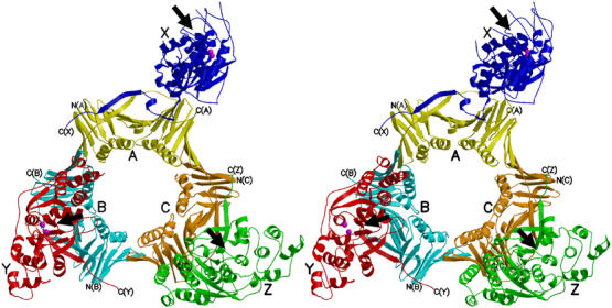
A stereo view of the human FEN1–PCNA complex. Three FEN1 molecules are colored in blue (X), red (Y) and green (Z), and the three subunits of the PCNA trimer in yellow (A), cyan (B) and orange (C). The C-termini of FEN1 and PCNA are labeled. Metal ions bound to the active sites of FEN1 (X and Y) are shown in magenta. Proposed catalytic faces of FEN1 are indicated by arrows.