
Fig. 3.
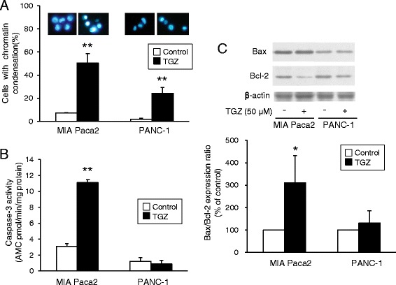
Apoptosis assays for TGZ. a Representative fluorescence microscopy images of cells stained with Hoechst 33342 and the percentage of cells with chromatin condensation. Cells were treated with TGZ (50 μM) for 24 h and stained with Hoechst 33342 for 15 min at 20 °C. Cells were then observed using fluorescence microscopy under UV excitation, and the percentage of cells showing chromatin condensation was determined. Data represent the mean + SD from four independent preparations. b Effects of TGZ on caspase-3 activity. Cells (6 × 10⁴ cells/well) were seeded in 24-well plates followed by 24 h incubation. After exposure to TGZ (50 μM) for 8 h, caspase-3 activity was assessed using a fluorometric Caspase 3 Assay Kit. Enzyme activities were determined as initial velocities corrected by protein quantity (n = 4). c Bax and Bcl-2 protein expression. Cells (1.75 × 10⁶) were incubated for 24 h and exposed to TGZ (50 μM) for another 24 h. Extracted protein (15 μg) was analyzed by western blotting. Bcl-2 expression levels were corrected using β-actin and analyzed from three independent preparations. * p < 0.05 and ** p < 0.01 vs. control (t-test). TGZ, troglitazone