Figure 1.

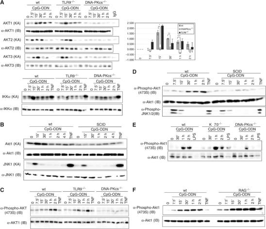
DNA-PKcs is involved in activation and phosphorylation of Akt in BMDMs. (A) Left panel: wt, DNA-PKcs−/− and TLR9−/− BMDMs were treated with CpG-ODN (10 μg/ml), TNF (10 ng/ml) or left untreated. At the indicated durations, cells were lysed by a lysis buffer (200 mM NaCl). A 200 μg portion of cell lysates was used to determine Akt KA by a kinase assay using GSK3α/β as a substrate (upper left panel) or IKK KA using GST-IκBα(1–54) as a substrate (lower left panel). The equal presence of samples was determined by an immunoblotting (IB) analysis using anti-Akt1, anti-Akt2 and anti-Akt3 antibodies. Right panel: quantitative analysis of Akt1 KA (relative fold) in wt, DNA-PKcs-deficient and TLR9-deficient BMDMs was performed using the Sigma Gel software. Data are mean±s.e.m. (n=4). *P<0.018, **P<0.003 and ***P<0.005 indicate statistical significance of Akt KAs in wt versus that in DNA-PKcs-deficient BMDMs treated by CpG-ODN for 7.5, 15, 30, 60 or 120 min. (B) wt and SCID BMDMs were treated with CpG-ODN (10 μg/ml), TNF (20 ng/ml) or left untreated. At the indicated durations, cell lysates were prepared. Akt KA was determined. JNK KA was measured by a kinase assay using GST-ATF2(6–96) as a substrate. The equal presence of proteins was determined by an IB analysis. (C–F) wt, DNA-PKcs−/−, TLR9−/−, SCID, Ku70−/− and Rag1−/− BMDMs were treated with CpG-ODN (10 μg/ml), LPS (3 μg/ml) or TNF (10 ng/ml) for the indicated durations or left untreated. Cell lysates were prepared and phosphorylation of Akt or JNK1/2 was detected using anti-phospho antibodies against Akt (473S) and JNK1/2, respectively.
