Figure 5.

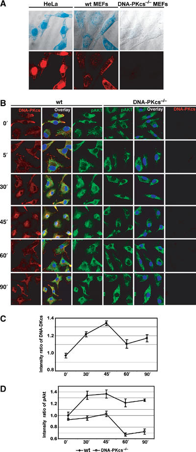
CpG-ODN induces DNA-PKcs and DNA-PKcs-dependent pAkt nuclear translocation. (A) Localization of DNA-PKcs in both HeLa and MEFs. HeLa, wt MEFs and DNAPKcs−/− MEFs were stained with anti-DNA-PKcs (mAb, cocktail) and anti-mouse-rhodamine antibodies. Images were detected by a confocal microscopy. The fluorescent images (lower panels) were superimposed on a bright field image of the respective cells (upper panels). (B) pAkt translocates into the nucleus in a DNA-PKcs-dependent manner following CpG-ODN treatment. wt and DNA-PKcs−/− BMDMs were treated with CpG-ODN (10 μg/ml) as indicated, fixed, permeabilized and immunostained with an anti-DNA-PKcs mAb (cocktail)/rhodamine and an anti-phospho-Akt (308T) antibody/FITC. The nuclear region was identified with DAPI staining. Quantitative analysis of DNA-PKcs (C) and pAkt (D) translocation was performed using the Leica Confocal software: the ratio of mean intensity of DNA-PKcs (C) or pAkt (D) signal in the nuclear region versus the total mean intensity for the respective cell was calculated for at least 10 randomly selected cells from several different fields. The means of all calculated ratios for DNA-PKcs in wt BMDMs (C) or pAkt in wt and DNA-PKcs−/− BMDMs (D) following CpG-ODN treatment were graphed. The nucleus was defined by DAPI staining.
