
Figure 5.
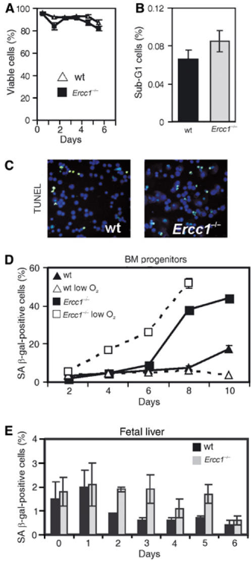
Decreased reserve capacity in the Ercc1−/− fetal liver erythroid progenitor pool is associated with progenitor senescence. (A) Fetal liver progenitor cultures were stained with the vital dye Trypan blue to determine viability at multiple time points after the initiation of cultures. A minimum of 200 cells were analyzed at each time point and the fraction of viable cells plotted. (B) BM progenitors were isolated from 21-d-old Ercc1−/− mice and wt littermates (three pools of two animals each), stained with propidium iodide to determine DNA content and analyzed by flow cytometry. Cells with less than 2N DNA content were gated and the percent calculated from the total number of viable cells. (C) TUNEL assay on fetal liver progenitors. Cells were isolated and cultured under growth-promoting conditions. Replica platings were harvested and analyzed for apoptosis every 24 h after isolation for a total of 8 d. No difference in the fraction of apoptotic cells between Ercc1−/− and wt cultures was detected. An example image from day 5 is depicted. (D) BM progenitors were isolated from femurs of 3-week-old Ercc1−/− mice and wt littermates (n=3, each genotype) and cultured for 10 d under growth-promoting conditions at either 20 or 3% (low) O2. A replicate of the culture was fixed every 2 d and stained for SA β-gal. Positively staining cells were counted and plotted as a percentage (mean±s.e.m.). (E) Fetal liver progenitors were isolated from three mice of each genotype and cultured as described in (A), then washed, fixed and stained for SA β-gal. The percentage of positively staining, senescent cells is plotted (mean±s.e.m.).