. 2015 Mar 31;6(7):3712–3717. doi: 10.1039/c4sc03900a

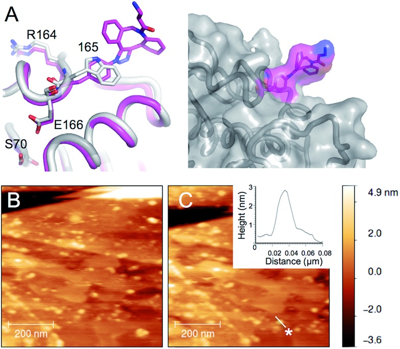
Fig. 4. AFM imaging of pyrene-modified TEMW165azF on graphene. (A) Model of TEMW165azF (magenta) modified with 1 aligned with the crystal structure of wt TEM (grey) with the residues either side of residue 165 and the catalytic S70 highlighted. The surface representation of TEMW165azF modified with 1 (left panel) illustrates that the Click adduct points out and away from the protein. Repeated AFM imaging of the surface after the (B) sixth and (C) tenth scans shows that the proteins bind stably to the surface. The height analysis of a single TEMW165azF-3 molecule, which is selected in C (highlighted as *) is shown as an inlay to C. ESI Movie 1 provides a full trace for the imaged area above over 10 scans.

Fig. 4