
Figure 8.
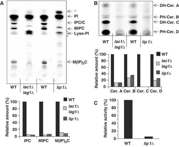
Labellings of Lip1Δ mutant. (A) Cells (strains RH4838, RH5308 and RH 6013) were labelled with [3H]myo inositol, lipids were analyzed on TLC and detected using a phosphorimager. Radioactivity in sphingolipids was quantified, and the relative amount of each species was determined as a percentage of the amount in WT cells. PI, phosphatidylinositol; IPC/C: inositolphosphorylceramide C; MIPC: mannose inositolphosphorylceramide; M(IP)2C: mannose di(inositolphosphoryl)ceramide; * indicates a spot that migrates identically to that previously identified as a C26 fatty acid containing PI. (B) The same cells were labelled with [3H]DHS and lipids were analyzed as above. Radioactivity in sphingolipids was quantified as in (A). (C) Ceramide synthesis activity of eluates from a Flag immunoisolation of digitonin-solubilized membranes from strains RH5666 and RH6075.