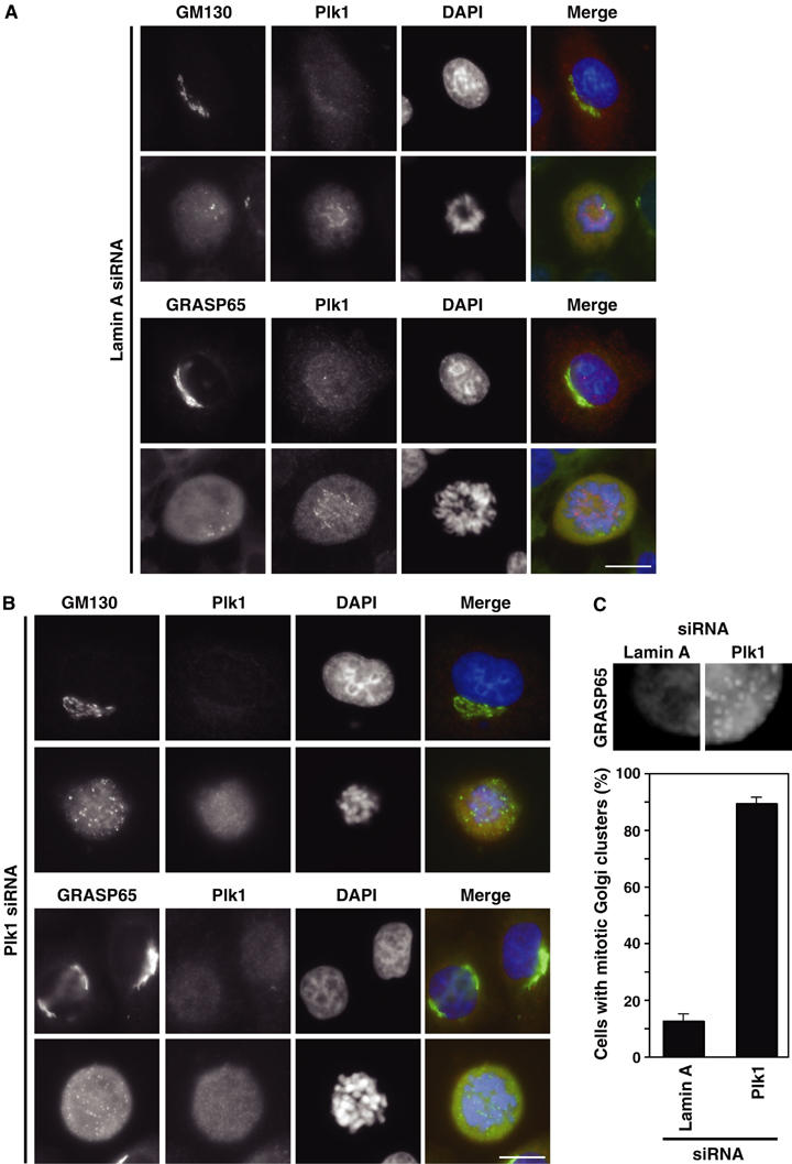
Figure 7.

Effect of Plk1 depletion on mitotic Golgi fragmentation. HeLa cells or HeLa cells stably expressing GRASP65-GFP were treated with siRNA duplexes for (A) lamin A and (B) Plk1 for 72 h, and then stained with antibodies to GM130 (green) and Plk1 (red), or stained with an antibody to Plk1 (red) and GRASP65 visualised using GFP (green). DNA was stained with DAPI (blue). Bar indicates 10 μm. (C) Enlarged images showing the increased number of persistent Golgi clusters, marked by GRASP65-GFP, observed in Plk1 siRNA cells. Numbers of mitotic cells containing Golgi clusters were counted for lamin A and Plk1 siRNA and plotted as a bar graph (300 cells per experiment, n=3).
