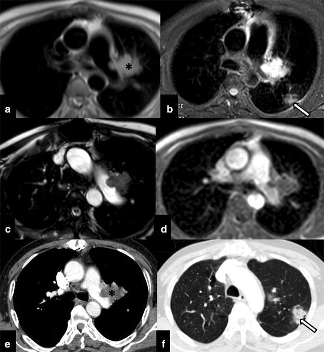
Fig. 3.

Post pulmonary endarterectomy cine-cardiac MRI (axial a, b, c, d T1W; b STIR, T2 and Post-contrast respectively) shows residual disease (asterisk) in the left pulmonary trunk with exophytic extravascular component. Focal patchy consolidation like opacities (arrow) were also seen in the lung parenchyma which were in favour of chronic thromboembolic phenomenon related infarcts presenting as consolidation (confirmed on post-contrast CT thorax: e mediastinal window and f lung window)
