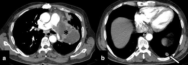
Fig. 4.

Follow-up imaging after 12 cycles of radiotherapy shows a large lobulated peripherally enhancing exophytic mediastinal mass (asterisk) arising from the left main pulmonary artery and invading its segmental arteries with a well-defined soft tissue nodule in left lower (arrow). CT findings were suggestive of disease progression with pulmonary parenchymal metastasis
