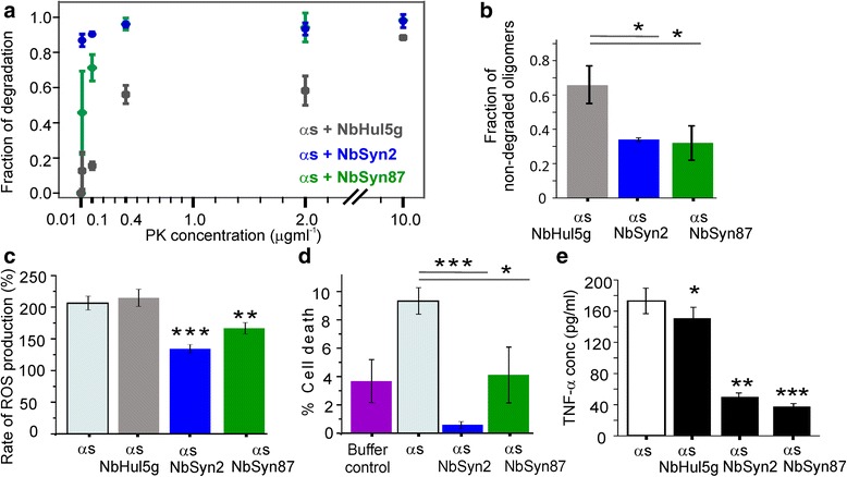
Fig. 4.

Comparative assays showing the relative stability and cytotoxicity of ɑS oligomers generated after aggregation for 29 h in the presence or absence of nanobodies under the same conditions as for the sm-FRET measurements. a Sm-FRET proteinase K digestion assays. The fraction of degradation is the number of oligomers in the proteinase K-exposed sample divided by the number of oligomers in the initial sample (n = 3, mean ± SD). Additional file 6: Figure S5 shows representative contour plots of FRET efficiencies versus apparent oligomer sizes. b Degradation of the 29 h samples by the 26S proteasome over 24 h (n = 3, SD, P < 0.05), analyzed by sm-FRET. c Cytoplasmic reactive oxygen species (ROS) production measured by monitoring the rate of dihydroethidium fluorescence (detailed in Additional file 2: Supplementary Methods). Application of ɑS oligomers to mixed cultures of rat hippocampal and cortical neurons (500 nM of total ɑS) induced an increase in the rate of ROS production relative to a control with no ɑS (taken as 100%); ɑS alone (n = 76 cells); ɑS with NbHul5g (n = 66 cells); ɑS with NbSyn2 (n = 89 cells, P < 0.001); and ɑS with NbSyn87 (n = 54 cells, P < 0.01). Additional file 8: Figure S6 shows representative plots of dihydroethidium fluorescence versus time. d Percentage cell death, measured by propidium iodide staining after incubation with the different ɑS oligomeric species. Cell death in the absence of ɑS (buffer control, n = 6 coverslips); upon incubation with ɑS alone (100 nM total ɑS, n = 7 coverslips); with ɑS with NbSyn2 (n = 5 coverslips, P < 0.001); with ɑS with NbSyn87 (n = 8 coverslips, P < 0.05). e Quantification of the released pro-inflammatory cytokine TNF-ɑ in BV2 microglia after the application of 29 h timepoints of ɑS (10 μM of the total ɑS), aggregated in the absence of nanobodies (n = 3, SEM), in the presence of 2 molar equivalents of NbHul5g (n = 3, SEM, P < 0.05), NbSyn2 (n = 3, SEM, P < 0.01), or NbSyn87 (n = 3, SEM, P < 0.001), and incubation with the cells for 24 h
