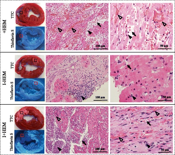
Fig. 6.

Injury Patterns on Histology. Images show TTC, Thioflavin S and H&E stained short axis sections obtained from explanted hearts at 24 h post intervention. TTC sections indicate region on necrosis appearing white in the I-HEM group (no hemorrhage) and reddish white in I+HEM group (hemorrhage); the +HEM group did no show infarction. Under ultra-violet light, non-fluorescent regions on the Thioflavin S stain highlight areas of compromised endothelium as seen in the +HEM and I+HEM groups; this confirms presence of microvascular damage in the hemorrhage groups. The H&E images were obtained from the region of interest shown on the TTC stained sections. Hemorrhage was apparent in the +HEM and I+HEM groups as evidenced from the interstitial distribution of red blood cells (open arrow heads) in the affected LAD territory; red blood cells were absent in the I-HEM group. Edematous development was observed in all three groups (arrows) along with the presence of inflammatory cells (closed arrow heads)
