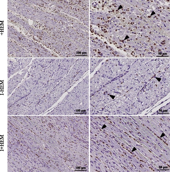
Fig. 7.

Macrophage Activity. Images show MAC387 macrophage stained sections from the LAD territory. Macrophage infiltration was extensive in both the hemorrhagic groups (+HEM and I+HEM) while it was scarce in the non-hemorrhagic infarct (I-HEM). Arrowheads indicate sites with macrophage activity. Notably, macrophages were predominantly concentrated beside the spilled red blood cells in the interstitial spaces. (Left column: 20x; Right column: 40x)
