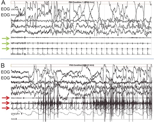
Figure 3. Polysomnographic (PSG) recordings.
PSG recordings of normal REM sleep (A) and REM sleep without atonia, typical of REM sleep behavior disorder (B).REM are reflected by the high-amplitude, abrupt deviations from baseline in the electro-oculogram (EOG) leads during a 30-second epoch. In (A), note the absence of EMG activity in the submental, leg, and arm leads (green arrows), whereas increased EMG tone is present in the same leads (red arrows) in B, particularly in the middle (arm lead), in this patient.

