Figure 1.
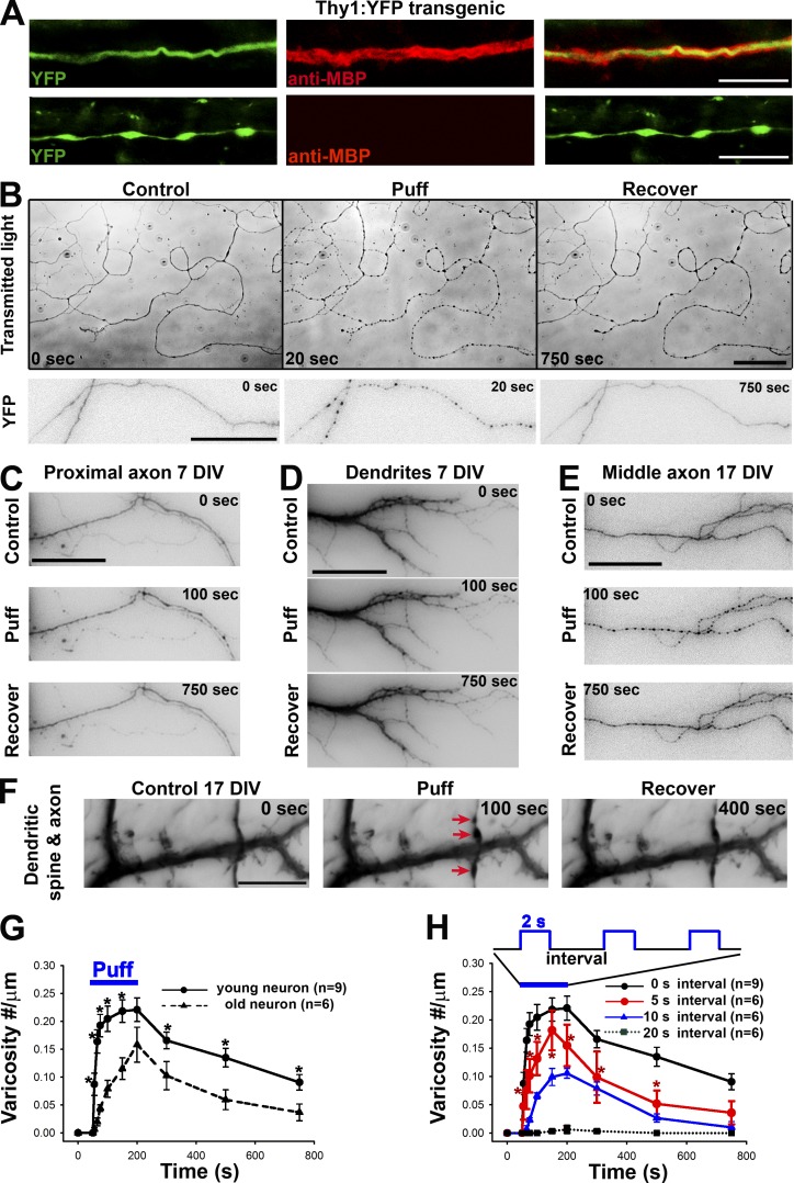
Mechanics-induced rapid and reversible initiation of axonal varicosities. (A) Axonal varicosities along unmyelinated (bottom) but not myelinated axons (top) in the hippocampus of Thy1-YFP transgenic mice. Axons are indicated by YFP fluorescence (green) and myelin segments by the anti-MBP staining (red). (B) Puffing induced rapid and reversible formation of axonal varicosities in cultured hippocampal neurons at 7 DIV, revealed by transmitted lights (top) and transiently expressed YFP (bottom). YFP signals are inverted. Transient or sustained mechanical pressures were delivered by puffing of Hank’s buffer via a glass pipette onto cultured hippocampal neurons as described in Fig. S1. The puffing time was 150 s. (C) Puffing did not induce varicosity formation in proximal axons (7 DIV). Neurons were transfected with YFP. The soma was on the left. (D) Puffing did not affect dendrite morphology (7 DIV). (E) Puffing induced varicosity formation in the middle axon of a neuron at 17 DIV. (F) Puffing induced varicosity formation along axons but not dendrites nor dendritic spines at 17 DIV. Red arrows indicate axonal varicosities. Related quantifications are included in Fig. S2. Bars: (A) 10 µm; (B) 50 µm; (C–F) 15 µm. (G) Older axons (21 DIV) were more resistant to puffing-induced varicosity formation than younger ones (7 DIV). (H) Frequency-dependent varicosity initiation in young axons receiving 2-s puffing pulses. Error bars show means ± SEM. Unpaired t test: *, P < 0.05. 5-s interval versus 20-s interval in H.

