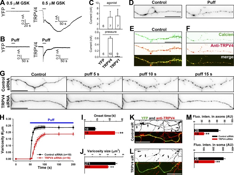
Figure 3.
TRPV4 channel plays a major role in axonal varicosity initiation in hippocampal neuron mechanosensation. (A and B) When expressed in HEK293 cells, TRPV4 but not YFP was activated by TRPV4 agonist GSK101 (GSK; 0.5 µM; A) and puffing (B). (C) Summary of the effects of their respective agonist (top) or puffing (bottom) on HEK293 cells expressing YFP, TRPV4, or TRPV1. (D) An axonal segment loaded with calcien AM (inverted signals) before (left) and after (right) puffing. (E) A distal axonal segment loaded with calcien AM (green) without puffing and stained for endogenous TRPV4 (red). (F) Post hoc staining of the axon in D for endogenous TRPV4 (red). (G) Effects of puffing on axons transfected with control (top) or TRPV4 (bottom) siRNA (siR). (H) The onset and degree of varicosity formation along axons with TRPV4 siRNA markedly reduced. (I and J) Under the same puffing condition, axons with TRPV4 siRNA had significantly longer onset time (I) and smaller varicosities (J). (K) Young axons (7 DIV) expressing YFP (green) and control siRNA were stained for endogenous TRPV4 (red in merged image and inverted in grayscale image). (L) Young axons (7 DIV) expressing YFP (green) and TRPV4 siRNA were stained for endogenous TRPV4 (red in merged image and inverted in grayscale image). Arrows indicate coexpressing YFP and double-stranded small RNA. Bars, 20 µm. (M) TRPV4 siRNA significantly reduced TRPV4 staining intensity in both axons and soma of cultured neurons. Error bars indicate means ± SEM. Unpaired t test: **, P < 0.01.

