Figure 1.
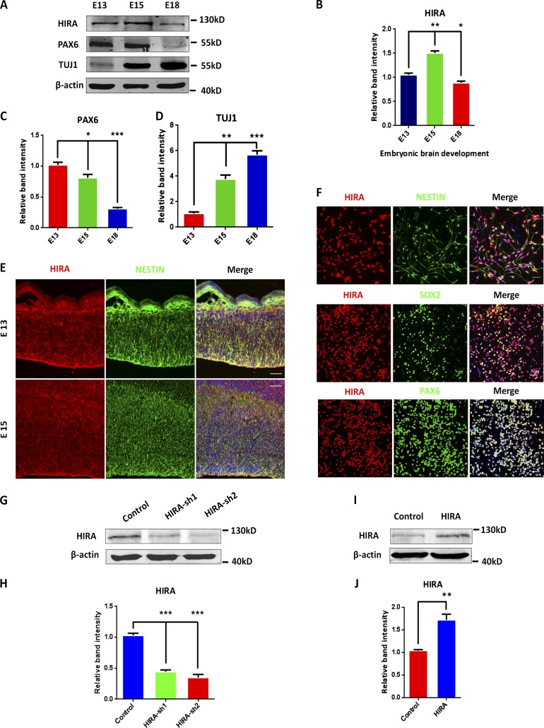
HIRA is expressed in the embryonic cerebral cortex and NPCs. (A) Western blot analysis of the protein levels of HIRA, PAX6, and TUJ1 in the mouse cerebral cortex during embryonic development. β-Actin was used as a control. (B–D) The bar graph displays the relative band intensity of HIRA (B), PAX6 (C), and TUJ1 (D) from embryonic day 13 to 18 (E13–E18; n = 3; mean ± SEM; *, P < 0.05; **, P < 0.01; ***, P < 0.001; t test, two sided). (E) E13 and E15 embryonic brain sections were costained with anti–HIRA and anti–NESTIN antibodies (VZ/SVZ). Bars: (E15) 25 µm; (E13) 50 µm. (F) NPCs were costained with anti–HIRA, anti–NESTIN, anti–SOX2, and anti–PAX6 antibodies. NPCs were isolated from E12.5 mouse brains and cultured in proliferation medium for 1 d. Bar, 25 µm. (G and H) In vitro–cultured NPCs were infected with control or HIRA shRNA lentivirus, and HIRA protein levels were analyzed using Western blot. The empty control shRNA was used as a control (n = 3; mean ± SEM; ***, P < 0.01; t test, two sided). (I and J) Western blot analysis shows the overexpression of HIRA in NPCs. The empty overexpression vector was used as a control (n = 3; mean ± SEM; **, P < 0.01; t test, two sided).

