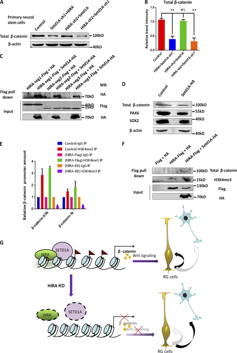
Figure 10.
Setd1A functions downstream of HIRA in regulating β-catenin–mediated NPC proliferation. (A and B) Western blot analysis of total β-catenin protein levels in NPCs infected with control, Setd1A-sh1 together with HIRA, HIRA-sh2 together with Setd1A, or HIRA-sh2 together with Setd1A-sh1 lentiviruses. The bar graph shows the relative band intensity of total β-catenin. β-Actin was used as a loading control (n = 3; mean ± SEM; **, P < 0.01; NS, not significant; t test, two sided). (C) HIRA segment 1, 2, or 3 all interact with Setd1A. The immunoprecipitated proteins were probed with anti–HA antibodies to detect HA-Setd1A and anti–Flag antibodies to detect Flag-HIRA-segment 1, 2, or 3. N2A cells were used in this experiment (n = 3). (D) Western blot analysis of the protein levels of PAX6, SOX2, and β-catenin in cultured NPCs infected with control or Setd1A-sh1 lentiviruses. β-Actin was used as a loading control (n = 3). (E) H3K4me3 deposition was increased at the β-catenin locus upon HIRA overexpression and was reduced at the β-catenin locus upon HIRA down-regulation. The anti–H3K4me3 antibody was used for immunoprecipitation (IP). The binding of H3K4me3 to the β-catenin promoter was determined through ChIP and real-time PCR (n = 3; mean ± SEM). (F) The expression levels of total β-catenin and H3K4me3 were increased in the anti–Flag antibody immunoprecipitated proteins when HIRA and Setd1A were coexpressed. N2A cells were used in this experiment (n = 3). (G) Model of HIRA function in neurogenesis during the embryonic development of the neocortex. HIRA inhibits neurogenesis by recruiting the H3K4 trimethyltransferase Setd1A to the β-catenin promoter. An increase of H3K4me3 expression promotes β-catenin expression and further enhances RGC proliferation. KD, knockdown; RG, retinal ganglion; WB, Western blot.

