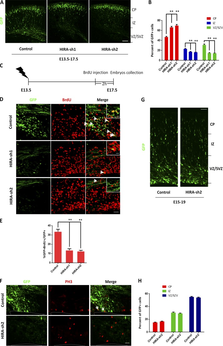
Figure 2.
HIRA regulates NPC distribution and proliferation. (A and B) HIRA knockdown changed GFP-positive cell distribution in the cortex. HIRA shRNAs or control plasmids were electroporated into E13.5 embryonic mouse brains, and embryos were sacrificed at E17.5 for phenotypic analysis. The percentage of GFP-positive cells in each region was analyzed (n = 3; mean ± SEM; *, P < 0.05; **, P < 0.01; t test, two sided). Bar, 50 µm. CP, cortical plate; IZ, intermediate zone; VZ/SVZ, ventricular zone/subventricular zone. (C–E) BrdU and GFP double-positive cells are reduced in HIRA shRNA plasmid–electroporated brains. Brains were electroporated at E13.5, and 100 mg/kg BrdU was injected i.p. into pregnant mice 2 h before the collection of embryos at E17.5. The arrows indicate GFP/BrdU double-positive cells. Insets show high-magnification view of control and HIRA-shRNA group. The bar graph displays the percentage of GFP/BrdU double-positive cells relative to the total number of GFP-positive cells in the VZ/SVZ (n = 3; mean ± SEM; **, P < 0.01; t test, two sided). Bars: (main) 25 µm; (insets) 10 µm. (F) The mitotic index of HIRA-silenced cells is decreased in utero. The percentage of PH3 and GFP double-positive cells in the ventricular zone is shown. Arrows indicate PH3 and GFP double-positive cells. Bar, 20 µm. (G and H) HIRA knockdown has no effect on cell migration. Control or HIRA shRNA plasmids were electroporated into E15 embryonic mouse brains, and embryos were sacrificed at E19 for phenotypic analysis. The percentage of GFP-positive cells in each region is shown (n = 3; mean ± SEM; t test, two sided). Bar, 50 µm.

