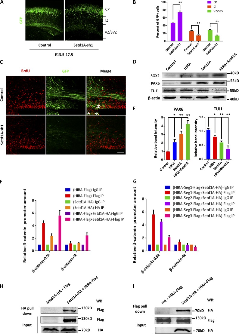
Figure 9.
The H3K4 trimethyltransferase Setd1A can cooperate with HIRA to modulate NPC proliferation. (A and B) Setd1A knockdown leads to changes in NPC distributions that are similar to that observed after HIRA loss of function. Setd1A-sh1 or control plasmids were electroporated into E13.5 embryonic mouse brains, and embryos were sacrificed at E17.5 for phenotypic analysis. The percentage of GFP-positive cells in each region is analyzed (n = 3; mean ± SEM; **, P < 0.01; t test, two sided). Bar, 50 µm. (C) BrdU and GFP double-positive cells are reduced in Setd1A-sh1 plasmid–electroporated brains. The brains were electroporated at E13.5, and BrdU (100 mg/kg i.p.) was injected into pregnant mice 2 h before the collection of embryos at E17.5. The arrows indicate GFP/BrdU double-positive cells. Bar, 25 µm. (D and E) Setd1A functions together with HIRA to modulate NPC proliferation in vitro. Cultured NPCs were infected with control vector, HIRA vector, Setd1A vector, or HIRA vector together with Setd1A vector, and the protein levels of PAX6, TUJ1, and SOX2 were analyzed using Western blot. The empty control expression vector was used as a control. The bar graph shows the relative band intensity of PAX6 and TUJ1. β-Actin was used as a loading control (n = 3; mean ± SEM; *, P < 0.05; **, P < 0.01; t test, two sided). (F) HIRA and Setd1A function together regulate β-catenin levels by binding to the β-catenin promoter. Primary NPCs were infected with control, HIRA overexpression, Setd1A overexpression, and HIRA together with Setd1A overexpression lentiviruses. HIRA and Setd1A binding to the β-catenin promoter was determined using ChIP and real-time PCR (n = 3; mean ± SEM). (G) HIRA-Segment 1 and Setd1A collectively modulates β-catenin levels by binding to the β-catenin promoter. Primary NPCs were infected with HIRA-Seg1-Flag, HIRA-Seg2-Flag, or HIRA-Seg3-Flag together with Setd1A-HA co-overexpression lentiviruses, and the anti–Flag antibody was used for immunoprecipitation. Protein binding to the β-catenin promoter was determined through ChIP and real-time PCR (n = 3; mean ± SEM). (H and I) HIRA and Setd1A can be immunoprecipitated together. The immunoprecipitated proteins were probed with anti–HA antibodies to detect HA-Setd1A and anti-Flag antibodies to detect Flag-HIRA. N2A cells were used in this experiment (n = 3). WB, Western blot.

