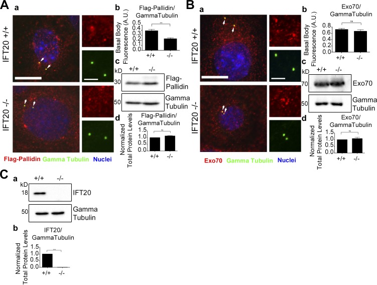
Figure 4.
Localization of pallidin at the basal body is dependent on IFT20. (A) Wild-type and Ift20−/− MEK cells expressing Flag-Pallidin. (Aa) Flag antibody staining (red), γ tubulin antibody staining (green), and nuclei detected with DAPI (blue). Flag-Pallidin localizes at the basal body (arrows). (Ab) Mean steady-state levels of Flag-Pallidin at the basal body are decreased in the Ift20-null cells. (Ac) Selected immunoblot images of total Flag-Pallidin and γ tubulin loading control protein levels. (Ad) No difference in the mean total protein levels of Flag-Pallidin in the Ift20−/− MEK cells compared with the control. (B) Endogenous Exo70 levels at the basal body in wild-type and Ift20−/− MEK cells. (Ba) MEK cells stained for endogenous Exo70 (red), γ tubulin (green), and nuclei detected with DAPI (blue). Endogenous Exo70 localizes at the basal body (arrows). (Bb) No difference in the mean steady-state levels of Exo70 at the basal body in the Ift20−/− cells compared with the control. (Bc) Selected immunoblot images of total endogenous Exo70 and γ tubulin loading control protein levels. (Bd) No difference in the mean total protein levels of Exo70 in the Ift20−/− cells compared with the control. (C) Total protein levels of endogenous IFT20 in wild-type and Ift20−/− MEK cells. (Ca) Selected immunoblot images of total endogenous IFT20 and γ tubulin loading control. (Cb) Mean immunoblot pixel intensity quantification showing no IFT20 protein present in the Ift20−/− MEK cells. n = 50 basal bodies per experimental group. Error bars are standard error of the mean. Data were analyzed using the unpaired Student’s t test. ***, P < 0.001; ****, P < 0.0001. Bars, 10 µm. Insets are 190% enlargements of the centrosome regions.

