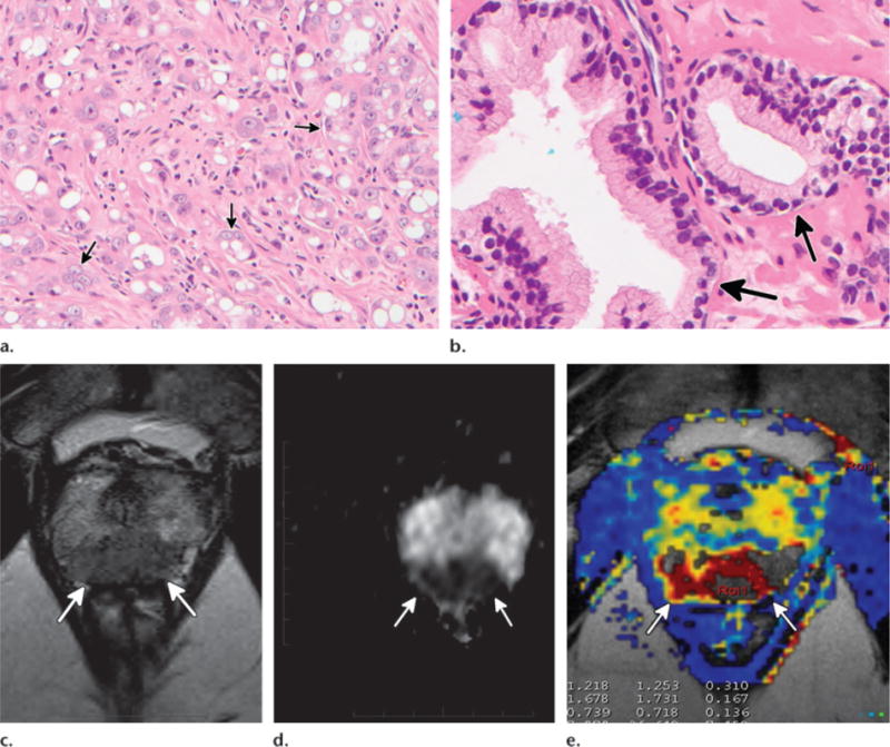
Figure 1.

Histologic and multiparametric MR imaging findings in prostate carcinoma. (a, b) High-power photomicrographs show poorly differentiated grade 5 prostate carcinoma with dispersed nests of carcinoma cells (arrows in a) and normal peripheral zone glandular tissue with well-formed glands (arrows in b). (Original magnification, ×200; hematoxylin-eosin [H-E] stain.) (c–e) Axial T2-weighted image (c), axial apparent diffusion coefficient (ADC) map (d), and axial ktrans image (e) show characteristic multiparametric MR imaging findings of high-grade (Gleason 8) prostate carcinoma in the midline apical peripheral zone (arrows).
