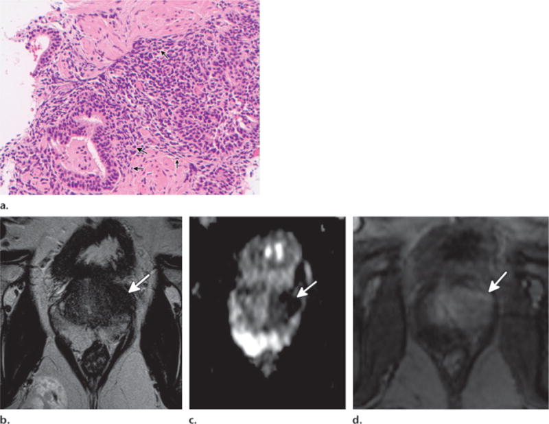
Figure 6.

Focal chronic prostatitis of the transition zone confirmed with MR imaging–guided biopsy in a 62-year-old man. (a) High-power photomicrograph of MR imaging–guided biopsy specimen shows extensive lymphocyte infiltration (arrows), in keeping with chronic prostatitis. (Original magnification, ×200; H-E stain.) (b–d) Axial T2-weighted image (b), axial ADC map (c), and axial DCE image (d) show a low-signal-intensity mass (arrow) in the left transition zone with diffusion restriction and early rapid enhancement.
