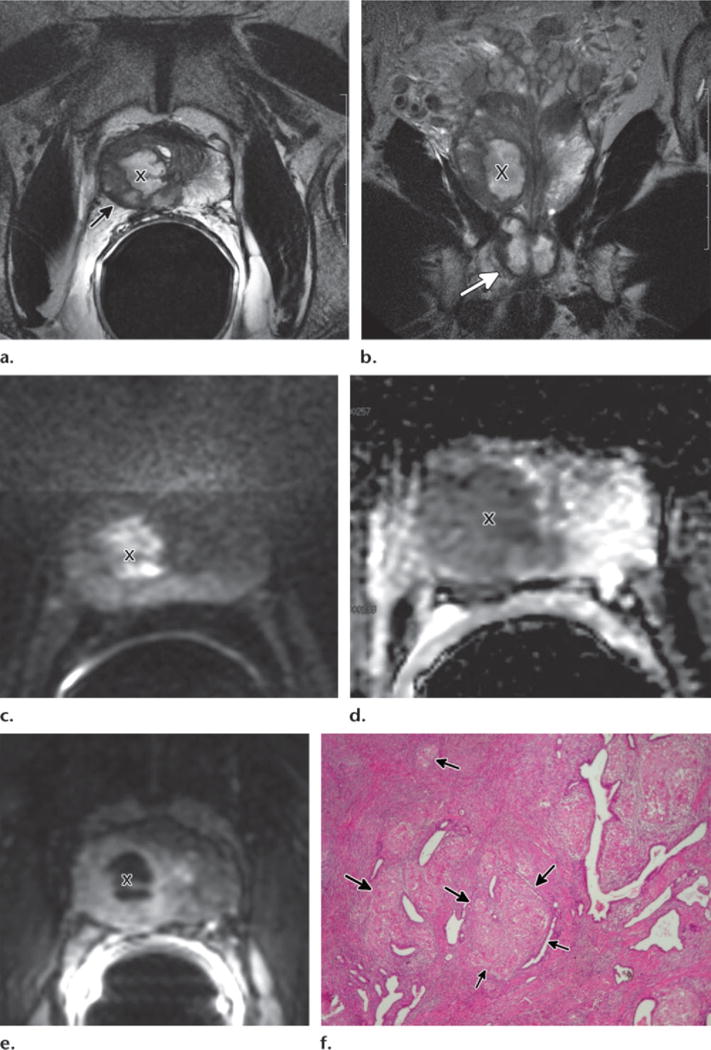
Figure 7.

Necrotic (caseating) mycobacterial granulomatous prostatitis and prostate cancer in a 64-year-old man with rising PSA level 2 years after completing intravesical bacillus Calmette-Guérin therapy. Transrectal US–guided biopsy revealed Gleason 6 prostate cancer and granulomatous prostatitis in the right lobe. (a–e) Axial T2-weighted image (a), coronal T2-weighted image (b), axial DW image (c), axial ADC map (d), and axial DCE image (e) show cavitating mycobacterial granulomatous prostatitis with extraprostatic extension. Cavitation (X) appears as high signal intensity on the T2-weighted images, hyperintensity on the high-b-value DW image, low ADC, and lack of enhancement. The abscess is surrounded by granulomatous infiltration, which appears as low T2 signal intensity, diffusion restriction, and moderate enhancement. The caseating process shows extraprostatic extension on the axial T2-weighted image with capsular irregularity and bulging (arrow in a). It also extends through the apex of the prostate (arrow in b) on the coronal T2-weighted image. (f) Low-power photomicrograph shows inflammation with granuloma formation (arrows). (Original magnification, ×40; H-E stain.)
