
Figure 3.
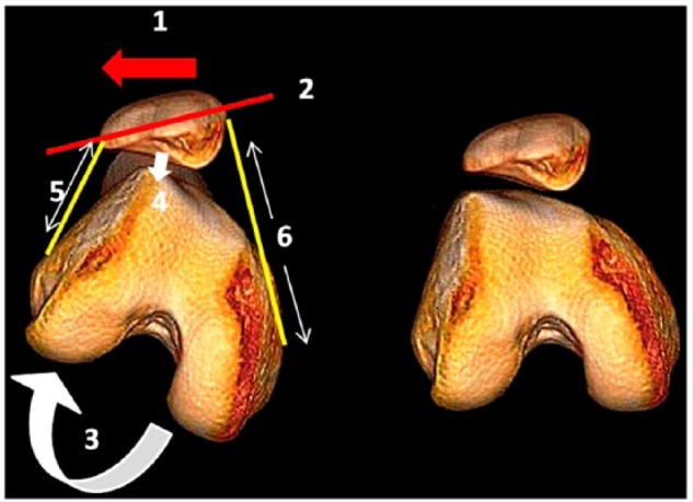
Three-dimensional computed tomography demonstrating abnormal patellar tracking secondary to femoral internal rotation. On the right is a normal knee. The left shows how the rotating movement of the femur underneath the patella in the transverse plane leads to abnormal patellar tracking (1, lateral patellar subluxation and 2, patellar tilt). The patella maintains a horizontal position while the femur internally rotates; therefore, the patellar subluxation is not the result of patella moving on the femur but of the result of the femur rotating underneath the patella. 3, inward twisting of the knee. 4 (arrow), compression in the lateral patellofemoral joint increases. 5, retracted lateral retinaculum. 6, tension increases in the medial retinaculum. The final result is patellofemoral imbalance.