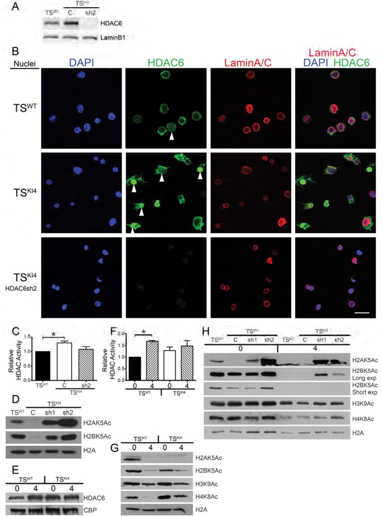
Figure 5. TSKI4 cells have increased nuclear HDAC6 localization and function.

(A) Nuclear HDAC6 protein levels are elevated in TSKI4 cells. Western blots of nuclear lysates are from TSWT or TSKI4 cells expressing a control shRNA (C) or TSKI4 cells expressing HDAC6 shRNA 2 (sh2).
(B) Immunofluorescence microscopy images of isolated nuclei show increased nuclear HDAC6 (green) in TSKI4 cells relative to TSWT cells. Nuclei were stained with DAPI (blue) and LaminA/C (red). Arrowheads indicate nuclear HDAC6. White bar represents 50 μm.
(A,B) Images shown are representative of three independent experiments.
(C) Increased total nuclear HDAC activity in TSKI4 cells expressed as the mean ± range of two independent experiments.
(D) Histone acetylation is restored by knockdown of HDAC6 in TSKI4 cells. Western blots are representative of three independent experiments.
(E–F) Differentiation and EMT induce nuclear HDAC6 localization and activity.
(E) Western blots of nuclear lysates from undifferentiated TSWT or TSKI4 cells (0) or cells differentiated four days (4). Blots are representative of two independent experiments.
(F) Nuclear HDAC activity assays were performed using nuclear lysates from undifferentiated cells or cells differentiated as in (E). Data are the mean ± range of two independent experiments.
(G–H) Western blots from cells undifferentiated (0) or differentiated for four days (4) as in (E). Blots are representative of three independent experiments.
(G) Histone acetylation is reduced with differentiation and EMT in TS cells.
(H) HDAC6 shRNA knockdown prevents differentiation-induced deacetylation of H2A/H2B.
Student’s t-test, *p-value < 0.05.
See also Figure S3.
