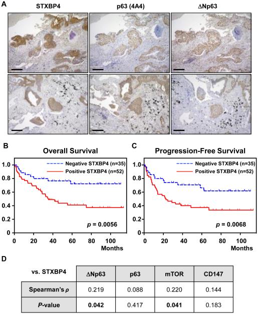
Figure 1.
STXBP4 expression is correlated with p63 expression and poor prognosis in lung SCC. (A) Representative immunohistochemical staining of a lung SCC. STXBP4 immunostaining demonstrates a nuclear and cytoplasmic pattern with a score of 5. Scale bars are 200 μm. (B, C) Kaplan-Meier analysis of Overall Survival (OS) and Progression Free Survival (PFS) defined according to STXBP4 expression. A statistically significant difference in OS and PFS was observed between the STXBP4-positive patients and those with low STXBP4 expression [OS, p = 0.0056(A); PFS, p = 0.0068 (B)]. P-values were obtained by log-rank test. (D) Spearman’s rank correlation was performed based on the expression levels of STXBP4 and ΔNp63.

