Figure 4.
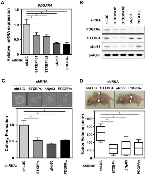
STXBP4-depletion inhibits lung SCC tumorigenesis and modulates PDGF signaling in vivo. (A) The lung SCC cells, RERF-LC-Sq1, were treated with siRNAs for Luciferase (siLUC) as a control, STXBP4#1, STXBP4#2, ΔNp63 or PDGFRα. Total RNAs were quantified by real-time RT-PCR analysis and the induction levels of PDGFRA were determined by the relative Ct method. (B) RERF-LC-Sq1 cells depleted as in (A), were subjected to immunoblotting using anti-PDGFRα, STXBP4, ΔNp63 or β-Actin antibodies. (C) The growth of RERF-LC-Sq1 cells after shRNA mediated PDGFRα, STXBP4 or ΔNp63 knockdown was monitored by soft agar colony formation assays. Standard deviations (SD) are plotted. *P < 0.05. (D) Representative images of xenografts from subcutaneously transplanted with lentivirally shRNA transduced Luciferase as a control (shLUC), Stxbp4, ΔNp63 or PDGFRα knockdown RERF-LC-Sq1 cells (n = 6 for each knockdown). The results of six independent injections of knockdown cells are shown. Twenty days after implantation, the length (L) and width (W) of the tumor mass were measured, and the tumor volume (TV) was calculated using the equation: TV = (L × W2)/2. *P < 0.05.

