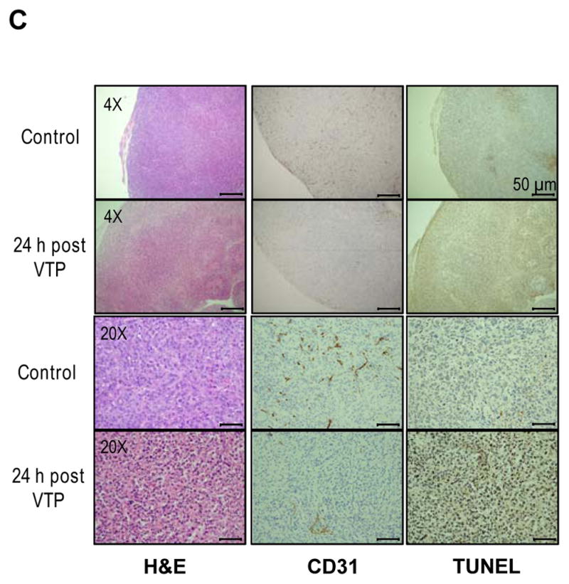
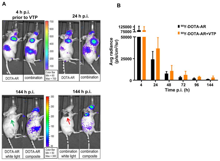
Figure 2. In vivo Cerenkov luminescence imaging (CLI) of 90Y-DOTA-AR in PC-3 tumor-bearing nude mice.

(A) Representative images of PC-3 xenografts treated with 90Y-DOTA-AR alone (DOTA-AR, n=12) or combination of 90Y-DOTA-AR and VTP (Combination, n=13) at 24 and 144 hours p.i. of 90Y-DOTA-AR. The images at 4 hours p.i. of 90Y-DOTA-AR show the uptake of 90Y-DOTA-AR prior to VTP treatment for both groups. After injection of 90Y-DOTA-AR, the radioactivity uptake within tumor was monitored using the IVIS Spectrum in vivo pre-clinical imaging system. Green arrows = tumor; red arrow = eschar formed post tumor ablation. (B) ROI analysis of 90Y-DOTA-AR CLI at each time point of imaging. VTP enhanced the retention of 90Y-DOTA-AR radioactivity within the tumors at all the time points measured after VTP treatment (p < 0.05). (C) PC-3 tumors were subjected to H&E staining and IHC of CD31 endothelial marker as well as TUNEL staining. Representative pictures of each group are shown. Tumor vasculature staining with anti-CD31 antibody was significantly reduced at 24 hours post VTP treatment alone compared to control specimen demonstrating VTP-induced tumor vessel destruction.

