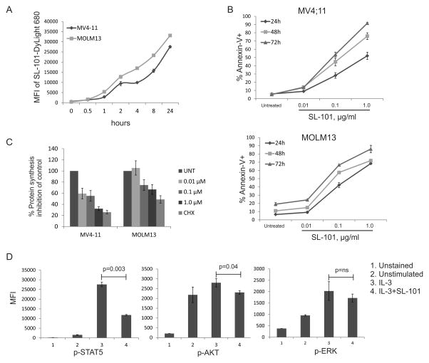
Figure 4. Mechanisms of the cytotoxic activity of SL-101.
(A) MOLM13 and MV4-11 cells were incubated (1 million/mL) with SL-101–DyLight 680 overnight, and internalized SL-101 was detected by flow cytometry on DyLight 680 signal after removing the membrane-bound SL-101. MFI, mean fluorescence intensity. (B) MOLM13 and MV4-11 cells were left untreated (untreat, or UNT) or treated with SL-101 at indicated concentrations for 24, 48, or 72 hours. Annexin-V+ apoptotic cells are shown. (C) MV4-11 and MOLM13 cells were treated with SL-101 (1 μg/mL) for 24 hours. Cycloheximide (100 μM) was used as a positive control. AHA (L-azidohomoalanine) reaction cocktails were applied to detect active protein synthesis. The signal was measured by a fluorescence microplate reader (485/520 nm) and the inhibition percentages were calculated in comparison with control group. (D) Mo7e cells (1 million/mL) were starved overnight and then stimulated with IL-3 (100 ng/mL) for 10 min. Cells were stained with antibodies after fixation and permeabilization.

