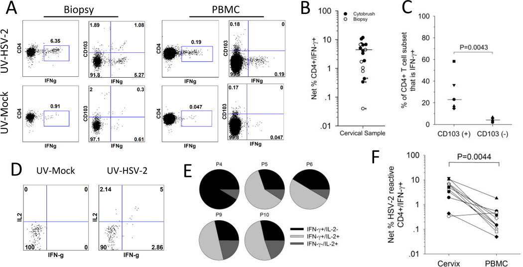
Figure 3. HSV-2 reactive CD4+ T cells are enriched in the CD103+ subset of cervical cells.
Cells isolated from the cervix or blood were co-incubated with UV-mock or UV-HSV-2 antigen and tested ex vivo for the expression of cytokines and CD103. (A) Representative dotplots comparing the reactivity of CD103+ and CD103− cells for IFN-γ expression in response to HSV-2 antigen. T cells from a cervical biopsy (left graphs) or PBMC (right graphs) from a representative participant were cultured with UV-mock (bottom graphs) or UV-HSV-2 (top graphs) and the resultant CD4+ T cells were analyzed by ICS and flow cytometry for the expression of IFN-γ and CD103. (B) Net % HSV-2 specific CD4+ T cells (CD4+/IFN-γ+) was measured in cytobrush and biopsy samples; *, proportion of CD4+/IFN-γ+ cells stimulated with UV-HSV-2 not statistically significantly different than those stimulated with mock (Fisher’s Exact test P>0.05). Median of all cervical samples where >100 cells analyzed is depicted by the bar. (C) HSV-2 reactive CD4+ T cells are enriched in the CD103(+) subset. The percentage contribution of IFN-γ+ (HSV-2 reactive) cells to the total CD103(+) and CD103(-) CD4+ T cell subsets from 5 participants is displayed. Bars represent the medians. (D) Representative dotplots displaying IFN-γ and IL-2 expression on CD4+ T cells derived from a cervical cytobrush sample from a representative participant co-incubated with mock (bottom graph) or UV-HSV-2 (top graph). (E) Co-expression of IFN-γ and IL-2 by HSV-2 reactive CD4+ T cells derived from cervical cytobrushes. The percentage of single and dual cytokine expressing HSV-2 reactive CD4+ T cells relative to the total HSV-2 reactive CD4+ T cell population is displayed. P, participant. (F) HSV-2 reactive CD4+ T cells are more abundant in the cervix compared to the blood. Net % HSV-2 reactive CD4+/IFN-γ+ T cells was calculated by ICS/flow cytometry from T cells derived from cervical cytobrushes (cervix) or PBMC.

